【衡道丨病例】乳腺低级别腺鳞癌(Low-grade adenosquamous carcinoma)如何诊断?
时间:2024-07-18 16:03:44 热度:37.1℃ 作者:网络
乳腺低级别腺鳞癌
定义
乳腺低级别腺鳞癌(Low-grade adenosquamous carcinoma)是一种罕见的乳腺化生性癌,具有腺、鳞状双向分化特征,细胞形态温和,并伴间质硬化的浸润性癌,预后较好。
临床特点
1.年龄:好发于中年女性,偶见于年轻女性。
2.部位:乳腺实质内可触及无痛性肿块,常位于外上象限。
形态学诊断要点
1.浸润生长模式
2.分化好的腺管
3.鳞状细胞巢团
4.梭形细胞背景
5.淋巴细胞聚集
6.可单发或伴发其他病变

瘤细胞在乳腺实质内杂乱无章地浸润性生长

腺腔圆形、拉长或者呈条索状(*所示)
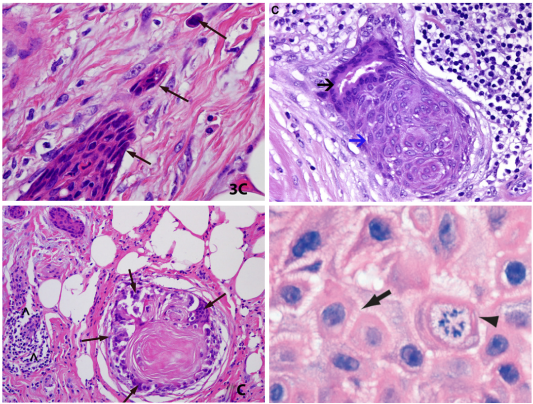
鳞状细胞巢可呈蝌蚪或逗点状,细胞分化好,可见角化珠和细胞间桥(箭头所指)

间质梭形细胞包绕上皮成分,细胞形态温和

常伴有淋巴细胞浸润

低级别腺鳞癌伴导管内乳头状瘤
免疫组化
1.ER、PR、HER2三阴性,AR阴性
2.肌上皮标记呈“完整、不连续、缺失”染色模式
3.上皮标记呈“核心染色”模式
4.间质梭形细胞表达SMMHC、Calponin,可呈“袖套样”染色模式
5.Ki67增殖指数低

SMMHC示肌上皮呈“完整连续、不连续、缺失”染色模式(箭头所指)

P63多样性表达(箭头所指):
①从完整、部分缺失到完全缺失的特征性改变;
②不管是鳞或腺的成分,都可以使瘤细胞核染色;
③可以使瘤细胞全部阳性,特别是鳞化的成分;
④可以表现为双层结构,腺上皮和周围的肌上皮。

CK示上皮呈“核心染色”模式

SMMHC示间质梭形细胞呈“袖套样”染色模式
分子改变
乳腺低级别腺鳞癌是一种低级别三阴性乳腺癌,具有基底样表型,AR阴性,PIK3CA 突变率高,无TP53 突变。
鉴别诊断
1、乳头部汗管瘤样肿瘤
(Syringomatous tumour of the nipple)
①发生于乳头部;
②可呈浸润性生长,会复发但不会转移;
③少见淋巴细胞聚集、间质细胞“袖套样”染色模式。

2、小管癌
(Tubular carcinoma)
①开放的小管,衬覆单层上皮细胞,常有胞突;
②一般不出现鳞状上皮化生,无淋巴细胞浸润;
③肌上皮标记物表达缺失;
④ER总是阳性,多数病例PR阳性。

3、硬化性腺病
(Sclerosing adenosis)
① 管状结构以小叶为中心分布,不呈浸润性的生长方式;
② 没有化生性成分;
③ 具有完整的肌上皮染色。

参考文献及书籍
1.WHO Classification of Tumours Editorial Board. WHO classification of tumours.Breast tumours[M]. 5th ed.Lyon: IARC Press, 2019:139- 154.
2.Senger JL, Meiers P, Kanthan R. Bilateral synchronous low-grade adenosquamous carcinoma of the breast: A Case report with review of the current literature. Int J Surg Case Rep. 2015;14:53-57.
3.Scali EP, Ali RH, Hayes M, Tyldesley S, Hassell P. Low-grade adenosquamous carcinoma of the breast: imaging and histopathologic characteristics of this rare disease. Can Assoc Radiol J. 2013;64(4):339-344.
4.Kawaguchi K, Shin SJ. Immunohistochemical staining characteristics of low-grade adenosquamous carcinoma of the breast. Am J Surg Pathol. 2012;36(7):1009-1020.
5.Bataillon G, Fuhrmann L, Girard E, et al. High rate of PIK3CA mutations but no TP53 mutations in low-grade adenosquamous carcinoma of the breast. Histopathology. 2018;73(2):273-283.
6.Boecker W, Stenman G, Loening T, et al. Differentiation and histogenesis of syringomatous tumour of the nipple and low-grade adenosquamous carcinoma: evidence for a common origin. Histopathology. 2014;65(1):9-23.

